Micro-CT Characterization of Abdominal Aortic Calcium Morphology
Abstract Body: Background: Calcium morphology is increasingly recognized as an important determinant of plaque biomechanics and procedural outcomes in coronary artery disease. In contrast, calcium morphology in the abdominal aorta (AA) remains poorly characterized, despite the AA being a common site of calcific disease and aneurysm formation. Furthermore, the limited spatial resolution of clinical imaging has constrained precise quantitative assessment of aortic calcium. In this study, we used non-clinical micro-computed tomography (micro-CT) to characterize abdominal aortic calcium morphology and its quantitative features.
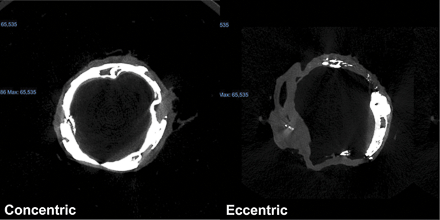
Methods: Abdominal aortic segments harvested between the renal arteries and aortic bifurcation from cadaveric donors (n = 10) were imaged using micro-CT. Cross-sectional analyses were performed throughout each segment, yielding 93 analyzed sections. Calcium morphology was classified at the section level as concentric or eccentric based on circumferential arc (concentric, >180 degrees). Quantitative measurements included calcium thickness, calcium area, calcium arc, and lumen area. Patient-level demographics and clinical characteristics (age, sex, smoking status, diabetes, cardiovascular disease, and other comorbidities) were compared across morphology classes, along with vessel-level quantitative parameters.
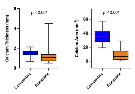
Results: Patient-level characteristics, including age, sex, smoking status, diabetes, and cardiovascular disease, did not differ between concentric and eccentric calcium groups (all p > 0.05). At the vessel-level, concentric calcium demonstrated significantly greater calcium thickness (median [IQR]: 1.46 [1.32, 1.71] mm vs. 1.06 [0.72, 1.38] mm, p < 0.001), and calcium area (34.3 [28.5, 41.9] mm^2 vs. 6.41 [2.24, 14.3] mm^2 p < 0.001) compared with eccentric calcium. Lumen area did not differ between morphologies.
Conclusions: Ultra-high-resolution non-clinical micro-CT enables detailed morphologic characterization of abdominal aortic calcification that is not achievable with conventional clinical imaging. While calcium morphology is increasingly incorporated into coronary assessment, its presence and implications in the abdominal aorta remain largely unexplored. Our findings demonstrate substantial morphologic heterogeneity in aortic calcium that is independent of systemic risk factors, suggesting a potential role for local biomechanical or biological drivers.
Elci, Gianna
(
New York Institute of Technology College Of Osteopathic Medicine
, Old Westbury , New York , United States )
Dharmasena, Pasani
(
New York Institute of Technology College Of Osteopathic Medicine
, Old Westbury , New York , United States )
Chong, Lionel
(
New York Institute of Technology College Of Osteopathic Medicine
, Old Westbury , New York , United States )
Bukhari, Zoraiz
(
New York Institute of Technology College Of Osteopathic Medicine
, Old Westbury , New York , United States )
Rahman, Sumona
(
New York Institute of Technology College Of Osteopathic Medicine
, Old Westbury , New York , United States )
Hurdle, Kelsi
(
New York Institute of Technology College Of Osteopathic Medicine
, Old Westbury , New York , United States )
Beatty, Brian
(
New York Institute of Technology College Of Osteopathic Medicine
, Old Westbury , New York , United States )
Author Disclosures:
Gianna Elci:DO NOT have relevant financial relationships
| Pasani Dharmasena:DO NOT have relevant financial relationships
| Lionel Chong:DO NOT have relevant financial relationships
| Zoraiz Bukhari:DO NOT have relevant financial relationships
| Sumona Rahman:DO NOT have relevant financial relationships
| Kelsi Hurdle:DO NOT have relevant financial relationships
| Brian Beatty:No Answer