Cardiac Imaging Uncovers Eosinophilic Vasculitis in a Patient With Pulmonary Embolism
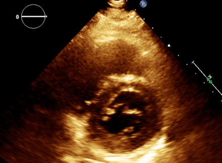
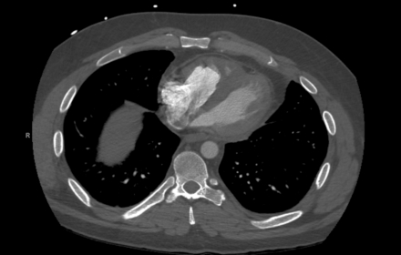
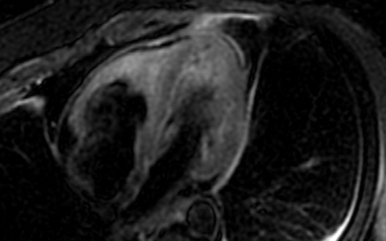
Abstract Body (Do not enter title and authors here): Description of case: A middle-aged man with a history of asthma and chronic sinusitis presented with pleuritic chest pain, breathlessness, and numbness in his left arm. On examination, he was hypertensive, tachypneic, and hypoxic. Laboratory workup showed leukocytosis with marked eosinophilia (13.09 x 10^3/μL), elevated cardiac biomarkers, and proteinuria. Computed tomography angiography revealed subsegmental pulmonary embolism. Echocardiography showed a moderately dilated right ventricle with reduced systolic function. Cardiac imaging identified diffuse myocardial edema, subendocardial enhancement in the right ventricle, and an apical thrombus. Cerebral magnetic resonance imaging revealed multifocal diffusion-restricted lesions. Coronary angiography showed no obstructive disease. Given the constellation of asthma, eosinophilia, neuropathy, and sinus disease, eosinophilic granulomatosis with polyangiitis (EGPA) was strongly considered. Testing for clonal eosinophilic syndromes and antineutrophil cytoplasmic antibodies was negative. Immunosuppressive therapy, including Rituximab and Mepolizumab, was initiated. Follow-up imaging at 9 months demonstrated complete resolution of myocardial inflammation and thrombus.
Discussion: Cardiac involvement in eosinophilic granulomatosis with polyangiitis is a major determinant of morbidity and mortality, contributing to nearly half of EGPA-related deaths. Clinical manifestations may include myocarditis, pericarditis, heart failure, or intracardiac thrombus. However, cardiac EGPA often lacks classic ischemic findings, making diagnosis challenging. Cardiac magnetic resonance imaging is the most sensitive modality for detecting myocardial inflammation, subendocardial fibrosis, and mural thrombus, particularly when endomyocardial biopsy is contraindicated. In this case, cardiac magnetic resonance imaging provided critical diagnostic clarity, revealing diffuse myocardial edema and subendocardial enhancement, features consistent with eosinophilic myocarditis. Integration of imaging findings with peripheral eosinophilia, asthma, neuropathy, and sinus disease enabled the timely recognition of EGPA. This case underscores the importance of early, multimodal cardiac imaging in patients with unexplained cardiac dysfunction and eosinophilia to prevent irreversible damage through targeted immunosuppression.
Kochhar, Gunjan
(
University of Oklahoma
, Oklahoma City , Oklahoma , United States )
Agrawal, Mukta
(
Oklahoma university
, Oklahoma city , Oklahoma , United States )
Pahuja, Mohit
(
University of Oklahoma COM
, Nichols Hills , Oklahoma , United States )
Author Disclosures:
Gunjan Kochhar:DO NOT have relevant financial relationships
| Mukta Agrawal:DO NOT have relevant financial relationships
| Mohit Pahuja:No Answer