Trends in left ventricular myocardial remodeling after thoracic endovascular aortic repair
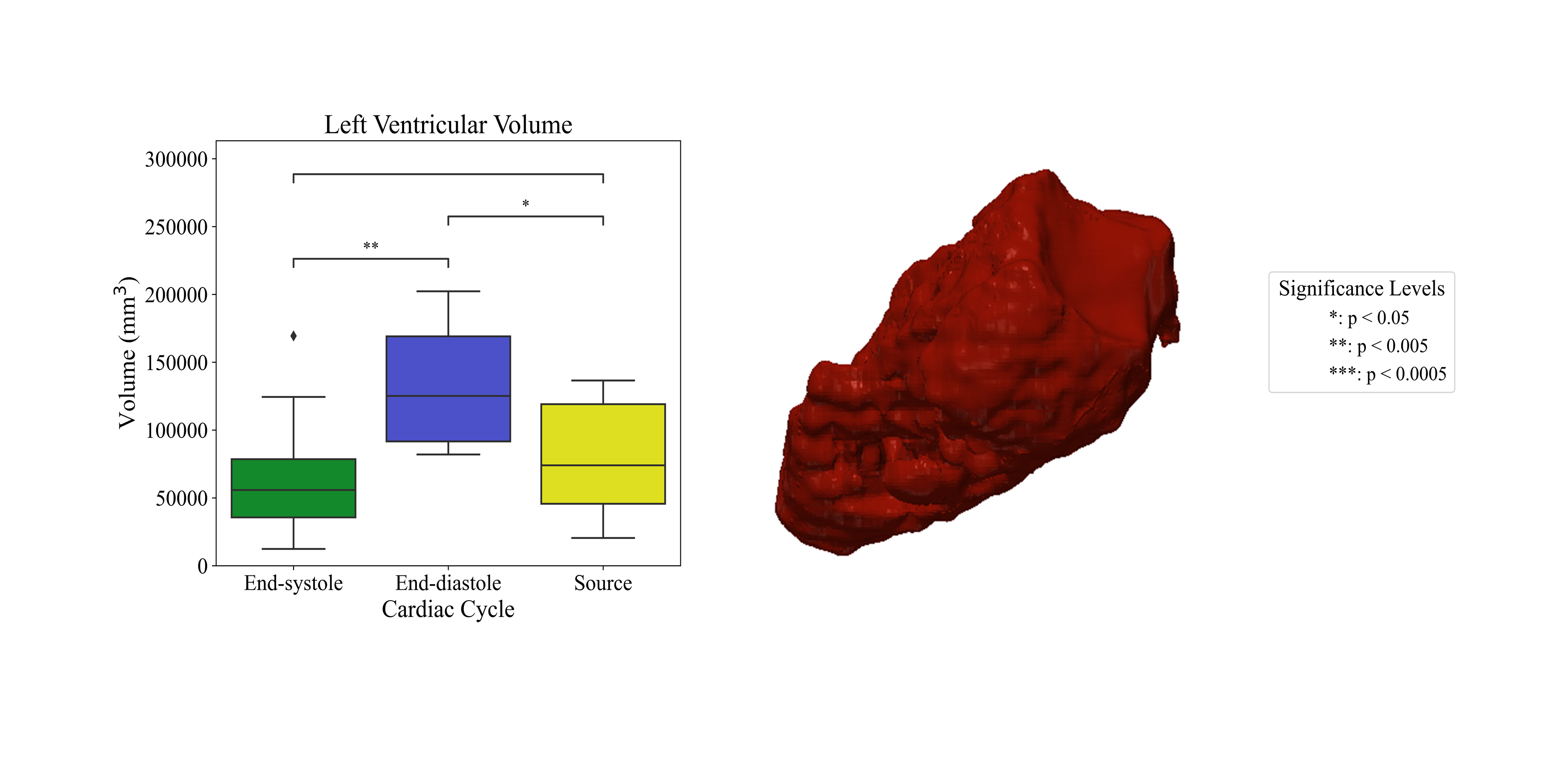
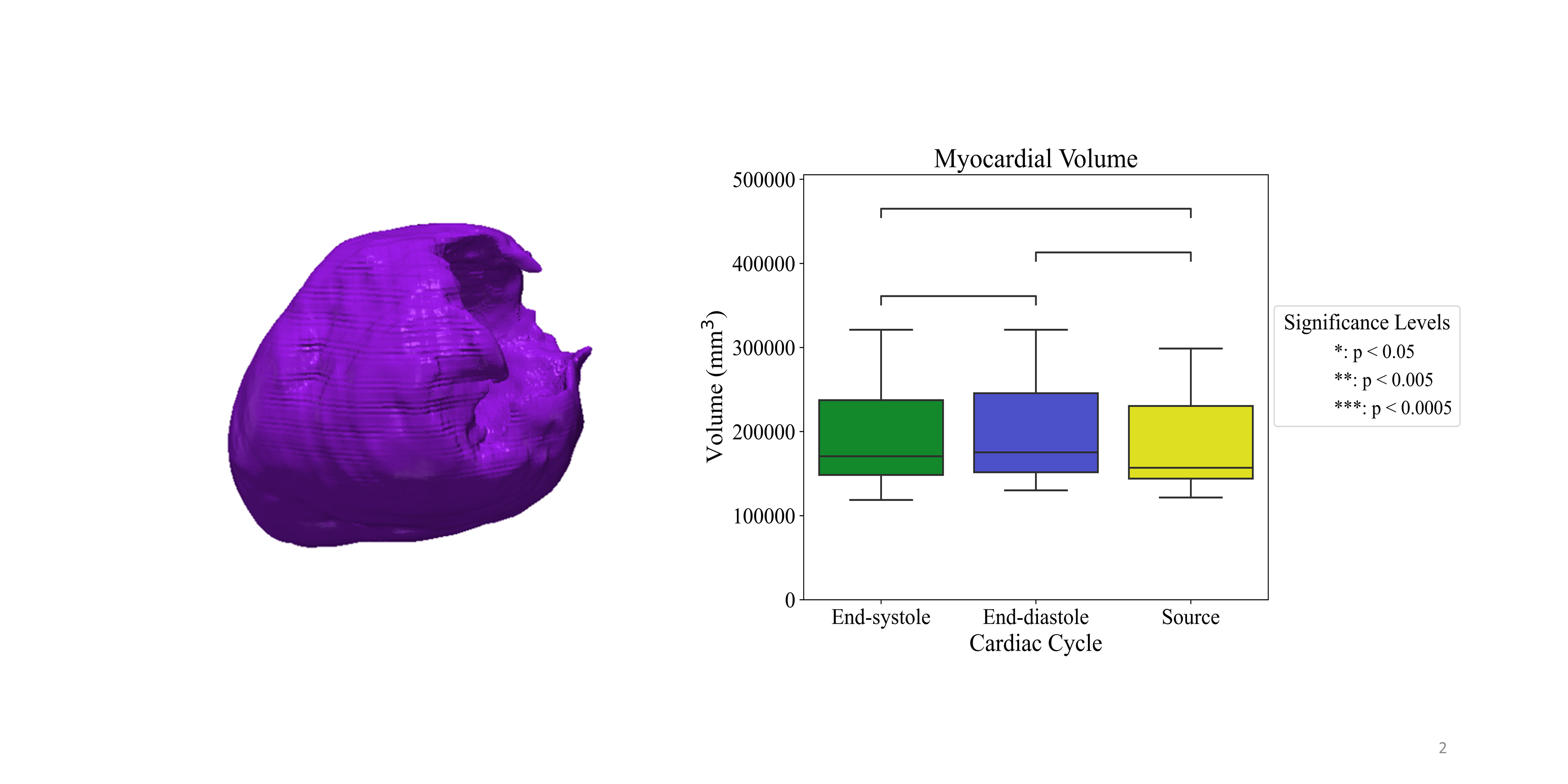
Abstract Body: Introduction: Thoracic endovascular aortic repair (TEVAR) has become a widely used treatment for various aortic pathologies. However, aortic endografts reduce aortic compliance and increase left ventricular afterload, potentially leading to arterial hypertension and heart failure. Further work needs to be done to elucidate the prevalence and etiology of cardiac disease after TEVAR implantation. This study aims to evaluate cardiac remodeling after TEVAR utilizing a large dataset of CTA scans from a diverse cohort of patients with aortic disease. Methods: To confirm preservation of left ventricular myocardial volume throughout the cardiac cycle, cardiac-gated CT scans from 11 patients were analyzed. Myocardium segmentations were generated automatically using Synopsys Simpleware ScanIP. These segmentations were performed at end-systole and end-diastole, as well as on the source CT. Two-dimensional surface meshes were created from these segmentations, and myocardial volumes were calculated. Student’s t-test was used to compare volumes across the cardiac cycle. Subsequently, a cohort of patients who underwent TEVAR was evaluated. A total of 1957 pre- and post-operative CT angiography (CTA) scans were analyzed. Indications for TEVAR included acute aortic dissection (92 patients), chronic aortic dissection (41 patients), traumatic aortic injury (96 patients), and thoracic aortic aneurysm (122 patients). For each scan, myocardial segmentation was performed automatically, a two-dimensional surface mesh was created, and myocardial volume was calculated. Results: Left ventricular myocardial volume was not significantly different at end-systole, end-diastole, and on the CT source image (190730 mm3 vs. 196571 mm3 vs. 189901.01 mm3, p=0.73). Preliminary data has been collected on 9 patients with chronic aortic dissection and 9 patients with thoracic aortic aneurysms. Among the dissection cohort, 5 of 9 patients (55.6%) exhibited post-TEVAR increases in myocardial volume, with a median increase of 3,275.2 mm3. Similarly, in the aneurysm cohort, 6 of 9 patients (66.7%) showed increases in myocardial volume post-TEVAR, with a median increase of 14,421 mm3. Conclusions: Because myocardial volume is preserved throughout the cardiac cycle, CTA imaging can reliably assess myocardial remodeling after TEVAR. Preliminary findings suggest TEVAR may be associated with increases in myocardial volume.
Twohig, Kelly
(
University of Chicago
, Chicago , Illinois , United States )
Jiang, David
(
University of Chicago
, Chicago , Illinois , United States )
Pugar, Joseph
(
University of Chicago
, Chicago , Illinois , United States )
Slivnick, Jeremy
(
University of Chicago
, Chicago , Illinois , United States )
Landeras, Luis
(
University of Chicago
, Chicago , Illinois , United States )
Pocivavsek, Luka
(
University of Chicago
, Chicago , Illinois , United States )
Author Disclosures:
Kelly Twohig:DO NOT have relevant financial relationships
| David Jiang:DO NOT have relevant financial relationships
| Joseph Pugar:DO NOT have relevant financial relationships
| Jeremy Slivnick:No Answer
| Luis Landeras:No Answer
| Luka Pocivavsek:No Answer