Long Duration of Type 2 Diabetes Promotes Erythrocyte-Induced Endothelial Dysfunction: Role of microRNA-210
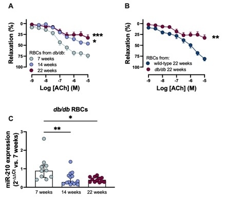
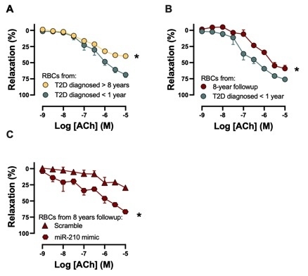
Abstract Body: Background: Type 2 diabetes (T2D) is a major risk factor for cardiovascular disease, with endothelial dysfunction playing a crucial role in the etiology of vascular complications. We previously demonstrated that red blood cells (RBCs) from T2D patients induce endothelial dysfunction via downregulation of miR-210 in RBCs. Notably, the duration of diabetes significantly increases the risk of cardiovascular disease. However, its influence on endothelial dysfunction induced by RBCs remains unexplored. Hypothesis: We test the hypothesis that long duration of T2D promotes the RBC-induced endothelial dysfunction, and this process is linked to miR-210 levels in RBCs. Methods: RBCs were isolated from wild-type mice (22 weeks), T2D db/db mice at different ages (7, 14, or 22 weeks) reflecting varying durations of disease, and humans with either newly diagnosed T2D (<1 year) or long-lasting T2D (>8 years). Aortas isolated from wild-type mice or Wistar rats were incubated with mouse and human RBCs, respectively. Following incubation, endothelium-dependent relaxation (EDR) was measured in the aortas using a wire myograph, and miR-210 levels were quantified in RBCs by qPCR. EDR was also re-evaluated in the newly diagnosed group after an 8-year follow-up. Results:db/db mice at all ages exhibited significantly elevated blood glucose levels compared to wild-type mice (mM: 26.5±4.1, 25.5±6.0, and 25.5±5.3 in 7-, 14-, and 22-week-old db/db vs.10.3±3.9 in 22-week-old wild-type mice). RBCs from 14- and 22-week-old db/db mice impaired EDR in wild-type mouse aortas, whereas EDR remained intact in vessels incubated with RBCs from 7-week-old db/db mice (Fig. 1A). RBCs from 22-week-old wild-type mice did not impair EDR (Fig. 1B). This suggests that T2D duration rather than aging per se contributes to endothelial dysfunction. Moreover, miR-210 levels in RBCs were significantly lower in 14- and 22-week-old db/db mice compared to 7-week-old db/db mice (Fig. 1C). In humans, RBCs from individuals with long-lasting T2D, but not newly diagnosed T2D, impaired EDR (Fig. 2A). Notably, after an 8-year follow-up of the newly diagnosed group, RBCs isolated from them impaired EDR (Fig. 2B), and this impairment was restored by the addition of a miR-210 mimic in RBCs (Fig. 2C). Conclusions: The duration of T2D appears to be a key factor in RBC-induced endothelial dysfunction, with this effect linked to reduced miR-210 levels in RBCs.
Kontidou, Eftychia
( Karolinska Institute
, Stockhokn
, Sweden
)
Zhou, Zhichao
( Karolinska Institutet
, Solna
, Sweden
)
Collado, Aida
( Karolinska Institute
, Stockhokn
, Sweden
)
Humoud, Rawan
( Karolinska Institute
, Stockhokn
, Sweden
)
Manickam, Kesavan
( Karolinska Institute
, Stockhokn
, Sweden
)
Jiao, Tong
( Karolinska Institute
, Stockhokn
, Sweden
)
Alvarsson, Michael
( Karolinska Institute
, Stockholm
, Sweden
)
Yang, Jiangning
( Karolinska Institute
, Stockhokn
, Sweden
)
Mahdi, Ali
( Karolinska Institute
, Stockhokn
, Sweden
)
Pernow, John
( Karolinska University Hospital
, Stockholm
, Sweden
)
Author Disclosures:
Eftychia Kontidou:DO NOT have relevant financial relationships
| Zhichao ZHOU:No Answer
| Aida Collado:DO NOT have relevant financial relationships
| Rawan Humoud:No Answer
| Kesavan Manickam:No Answer
| Tong Jiao:No Answer
| Michael Alvarsson:No Answer
| Jiangning Yang:DO NOT have relevant financial relationships
| Ali Mahdi:No Answer
| John Pernow:No Answer