Gap Junction Communication Facilitates Perivascular Adipose Tissue Function; Loss in Hypertension?
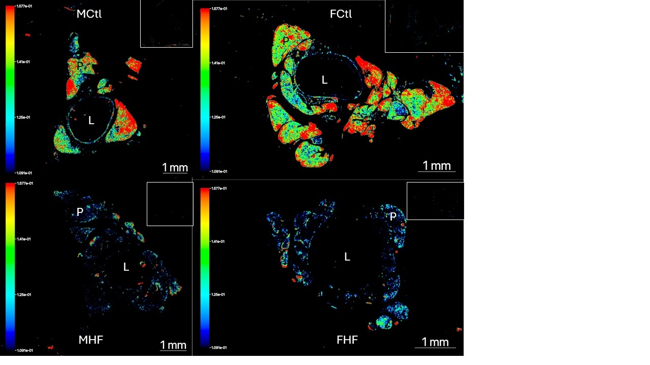
Abstract Body: Perivascular adipose tissue (PVAT) is not innervated and, here, we questioned how the cells of PVAT communicate to execute their functions. We hypothesize that gap junctional intercellular communication (GJIC) is one mechanism by which this occurs and focused on the function of stress relaxation. The thoracic aorta of the Dahl SS M and F rat was used. Aorta were from animals fed a control (10%) diet or high fat (60%) diet fed from weaning than induced a hypertension. The gap junction monomeric connexins were measured/visualized using snRNA sequencing, RNA scope, and immunohistochemistry. Isolated PVAT for measurement of isometric tone. In snRNA seq of the thoracic aortic PVAT (all rats), connexin 43 (Cx43; GJA1) was by far the most highly expressed connexin, present in adipocyte, endothelial cell, mesothelial cell, lymphatic endothelial cell, fibroblast but not immune cell (T cells, B cells, Macrophages) clusters. In the thoracic aorta, RNAscope supported expression of Cx43 in PVAT to a greater extent than the media. Similarly, Cx43 protein was located to the PVAT > aortic media (NovaRed visualized, brightfield). Importantly, Cx43 mRNA GJA1 in PVAT was 15-fold higher vs arterial media in the human epigastric artery (N=5, p=.003). In considering function, the stress relaxation of isolated PVAT, established as a mechanoresponse to stretch, was significantly reduced by the non-connexin specific inhibitor heptanol [mg relaxed to cumulative 8 gr stretch = 1337+112 mg; heptanol (1 mM): 2348+216 mg; p<0.05]. In the HF-fed Dahl SS rat, the potential reduction of GJIC in the condition of hypertension is supported by the finding that Cx43 protein expression was reduced in the PVAT of the aorta from HF vs control (immunohistochemically detected by Nova Red pseudo-colored at 800 nm on Odyssey M, arbitrary units/area PVAT; Control (M+F, N=10) = 3.91+0.35; HF (N=9) = 2.33+0.25, p<0.05). We conclude that gap junctions enable PVAT to function as a tissue, coordinating a response such as stress relaxation. The loss of Cx43 in hypertension supports a potential communication isolation of PVAT in disease.
Grabowski, Victor
(
Michigan State University
, East Lansing , Michigan , United States )
Lockwood, Lizbeth
(
Michigan State University
, East Lansing , Michigan , United States )
Thompson, Janice
(
Michigan State University
, East Lansing , Michigan , United States )
Contreras, Andres
(
Michigan State University
, East Lansing , Michigan , United States )
Wabel, Emma
(
Michigan State University
, East Lansing , Michigan , United States )
Lauver, Adam
(
Michigan State University
, East Lansing , Michigan , United States )
Terrian, Leah
(
Michigan State University
, East Lansing , Michigan , United States )
Nault, Rance
(
Michigan State University
, East Lansing , Michigan , United States )
Kung, Theodore A
(
The University of Michigan
, Ann Arbor , Michigan , United States )
Watts, Stephanie
(
Michigan State University
, East Lansing , Michigan , United States )
Author Disclosures:
Victor Grabowski:DO NOT have relevant financial relationships
| Stephanie Watts:DO NOT have relevant financial relationships
| Lizbeth Lockwood:DO NOT have relevant financial relationships
| Janice Thompson:DO NOT have relevant financial relationships
| Andres Contreras:DO NOT have relevant financial relationships
| Emma Wabel:DO NOT have relevant financial relationships
| Adam Lauver:DO NOT have relevant financial relationships
| Leah Terrian:DO NOT have relevant financial relationships
| Rance Nault:No Answer
| Theodore A Kung:No Answer
Ali Manzer, Umar Haddaya, Nazir Tahira, Nizam Muhammad, Steafo Lark, Sharif Ayesha, Jehangir Hanzala, Arham Muhammad, Hamza Anfal, Hassan Arbaz, Amjad Ans, Ali Iman, Zuha Zuha