Final ID: P-455
Integrins play a role in stress relaxation of perivascular adipose tissue
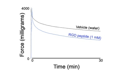
Abstract Body: Perivascular adipose tissue (PVAT) is well known for being anti-contractile (reducing potency, maximum) in healthy tissues. We discovered a new function of PVAT, the ability to stress relax in response to a stretch. This is of note because stress relaxation has been attributed largely to smooth muscle, of which PVAT has none that is organized in a functional layer. Here we test the hypothesis that stress relaxation depends on the interactions of the cell membrane delimited heterodimer integrins with collagen. Our model is the thoracic aorta of the male Dahl SS rat on a normal diet. The PVAT and aorta were physically separated for most assays. Results from snRNA seq in PVAT, histochemistry and isometric contractility were used. Collagen was evident in PVAT as stained by Masson Trichrome. snRNA seq of whole PVAT determined expression of many collagen isoforms and integrins, with mRNA for both the α1 and β1 integrin being most highly expressed. Isolated aortic rings and separated PVAT were challenged with 2 or 4 grams of passive tension. Pharmacological inhibition of integrin/collagen interaction was effected by the specific α1β1 distintegrin obtustatin (5 uM) or general inhibitor RGD peptide (1 mM). RGD peptide but not obtustatin reduced the ability of the isolated PVAT ring to maintain applied tone at both the 2 and 4 gram challenge (~15% gain in relaxation/loss of tone, figure). By contrast, stress relaxation in the aorta itself was not reduced by either inhibitor. In addition to a1b1 and collagen interaction, cell-cell communication inference identified integrins av and a5, two major RGD motif containing isoforms, as potential partners of collagens with fibroblast-like cells most likely involved. Collectively, these findings validate that stress relaxation can occur in a non-smooth muscle tissue, doing so at least in part through integrin-collagen interactions that may not include α1β1 heterodimers. The importance of this lies in considering PVAT as a vascular layer that possesses mechanical functions.
More abstracts on this topic:
Lineage-specific iPSC derived smooth muscle cells predict mannose receptor C type 2 deficiency accelerates aortic root enlargement in Marfan syndrome mice via exacerbating collagen deposition
Kusadokoro Sho, Yamaguchi Atsushi, Fischbein Michael, Yokoyama Nobu, Kim Jennifer, Bolar Nikhita, Dalal Alex, Pedroza Albert, Gilles Casey, Puaala Anna Marie, Klinder Avani
Effectiveness of Elective Endovascular Intervention for Patients with Peripheral Arterial Disease and Intermittent ClaudicationDhruva Sanket, Murillo Jaime, Ameli Omid, Conte Michael, Redberg Rita, Cohen Ken

