AAV cTnI gene therapy reverses diastolic dysfunction and derepresses fetal troponin I expression levels in juvenile cTnI null mice
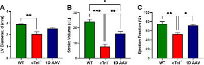
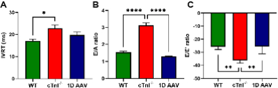
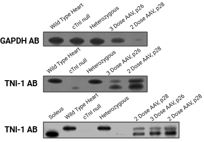
Abstract Body: Background: Mutations and loss of cardiac troponin I (cTnI) are often linked to diastolic dysfunction in the heart. We have developed a unique cTnI knockout mouse model, where homozygote cTnI null (cTnI-/-) mice survive past birth due to a fetal isoform of cTnI, identical to slow skeletal troponin I (ssTnI), but die at postnatal day (p) 17-18 from its progressive loss. In our previous study we investigated different dosing of adeno-associated virus serotype 9 (AAV9) carrying cardiac specific promoter (chicken cTnT) with wild type cTnI gene in cTnI-/- mice and showed 2-3 week extension of life and cTnI restoration. Interestingly, the current study indicates that gene therapy in cTnI-/- mice not only restores cTnI but results in a derepression of ssTnI. In depth echocardiography analysis was performed to examine cardiac function, and protein analysis was performed using Western blotting. Objectives: We tested the hypothesis that cTnI gene therapy will rectify the pathological defects in the myocardium under conditions of cTnI knockout (both at the level of echocardiography parameters and in terms of protein isoform expression). Methods: cTnI-/- mice were injected intraperitoneally with 5 x 10¹⁰ vg/g (vector genome per gram) AAV9. AAV administrations were as follows: single dose injection at p10, two-dose multi-injection at p10 & p14, and three-dose multi-injection at p10, p14, & p16. Cardiac function of AAV treated mice were assessed using two-dimensional B-mode and anatomical M-mode imaging and Doppler echocardiography at p18, single dose administration, and p25, single and multi-injection dosing. After heart failure, ssTnI and cTnI concentration in the heart were determined with Western blot analysis. Results: The data indicates that after 1 dose of AAV treatment, cTnI-/- mice at p18 showed partial and complete restoration of diastolic parameters including left ventricular diameter at end diastole (Figure 1A), stroke volume (Figure 1B), ejection fraction (Figure 1C), isovolumetric relaxation time (Figure 2A), E/A (Figure 2B), and E/E’ (Figure 2C). Protein analysis of two and three-dose multi-injection indicates the endogenous presence of the ssTnI isoform at significant levels (Figure 3). Conclusion: Administration of the cTnI gene therapy not only augmented levels of cTnI but also elicits compensatory mechanism for derepression of ssTnI. cTnI gene therapy, to a significant degree, reversed the indices of diastolic dysfunction.
Gerber, Dustin
(
Florida Atlantic University
, Sunrise , Florida , United States )
Hackshaw, Andrue
(
Florida Atlantic University
, Sunrise , Florida , United States )
Alexander, Joseph
(
Florida Atlantic University
, Sunrise , Florida , United States )
Cerqueira, Jessica
(
Florida Atlantic University
, Sunrise , Florida , United States )
Machi, Jacqueline
(
University of Miami
, Miami , Florida , United States )
Prentice, Howard
(
Florida Atlantic University
, Sunrise , Florida , United States )
Huang, Xupei
(
Florida Atlantic University
, Sunrise , Florida , United States )