Contrast-free ultrasound perfusion estimation and imaging of the calf muscle shows promise for peripheral arterial disease diagnosis
Abstract Body (Do not enter title and authors here): Introduction/Background Peripheral arterial disease (PAD) exhibits reduced hyperemic response in the affected legs following stimuli such as pressure-cuff-induced occlusion or exercise. Therefore, an analysis of the perfusion changes in the calf muscle using a widely available imaging modality such as ultrasound could serve as a complementary tool to existing methods such as ankle-brachial index (ABI) for PAD detection.
Research Questions/Hypothesis This study evaluates the utility of ultrasound perfusion imaging and estimation technique in the calf muscle to differentiate PAD affected legs based on their flow response to external pressure and exercise.
Methods/Approach Data acquisition was performed using a research ultrasound system (Vantage NXT, Verasonics) with an ultrasound probe secured inside a probe fixation device and positioned to the calf muscle of the subject. Two separate studies were performed on each subject: 1) A flow variations response to external pressure study, 2) a flow variation response to exercise. For the pressure study, a pressure cuff was wrapped around the subject’s thigh and inflated during the study using an automatic rapid cuff inflation device. Data was continuously acquired for 6 minutes including 1 minute of baseline, 3 minutes of pressure cuff occlusion, 2 minutes of post-occlusion acquisition. The exercise study consisted of data acquisition in the seated position with 1 minute of baseline, 30 repetitions of plantar flexion using a calf training device, and 2 minutes of post-exercise acquisition. The acquired data were then clutter-filtered and denoised for perfusion visualization. Four metrics including post-occlusion/post-exercise to baseline flow intensity/density variation (POBFIV, PEBFIV, POBFDV, PEBFDV) were obtained for analysis.
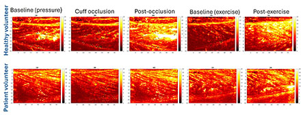
Results/Data Figure 1 shows perfusion imaging results during the pressure and the exercise studies for two example legs, one healthy and one of a clinically diagnosed PAD patient (mild disease), respectively. Percentage-wise POBFIV, PEBFIV, POBFDV, and PEBFDV values were 44%, 57%, 197%, and 2231% for the healthy leg and 12%, 27%, 63%, and 181% for the PAD leg, respectively, shown in Table 1.
Conclusion The proposed method shows promise in differentiating PAD-affected legs from healthy individual’s through evaluating the hyperemic response to external pressure and exercise. Statistical analysis of the results for a cohort of healthy and PAD-affected legs will constitute future work.
Sabeti, Soroosh
(
Mayo Clinic
, Rochester , Minnesota , United States )
Ahadi, Mahsa
(
Mayo Clinic
, Rochester , Minnesota , United States )
Mcbane, Robert
(
Mayo Clinic
, Rochester , Minnesota , United States )
Fatemi, Mostafa
(
Mayo Clinic
, Rochester , Minnesota , United States )
Alizad, Azra
(
Mayo Clinic
, Rochester , Minnesota , United States )
Author Disclosures:
Soroosh Sabeti:DO NOT have relevant financial relationships
| Mahsa Ahadi:No Answer
| Robert McBane:DO NOT have relevant financial relationships
| Mostafa Fatemi:No Answer
| Azra Alizad:No Answer