Comparison of the association of vascular aging constructed by different indices with the risk of cardiovascular events
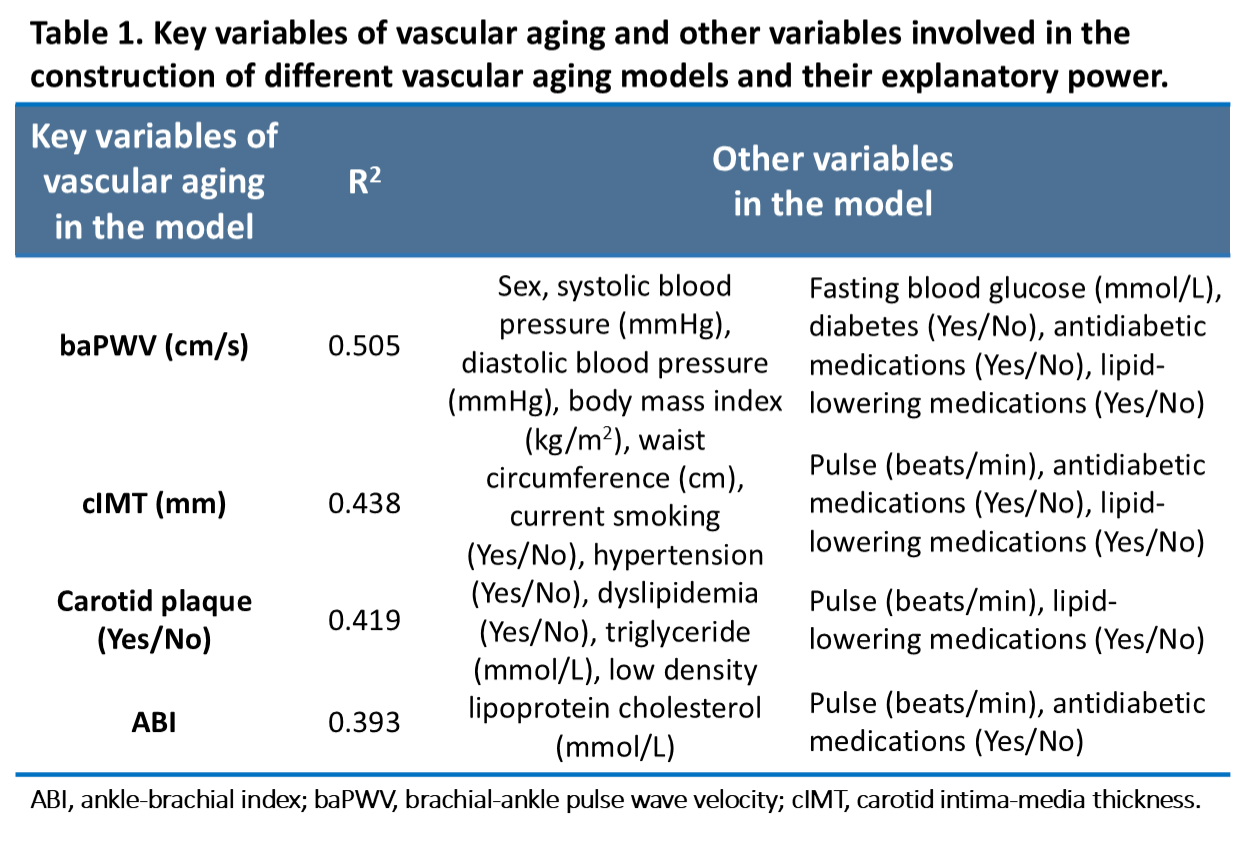
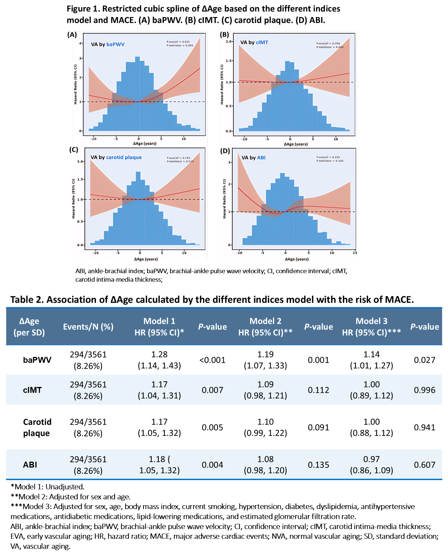
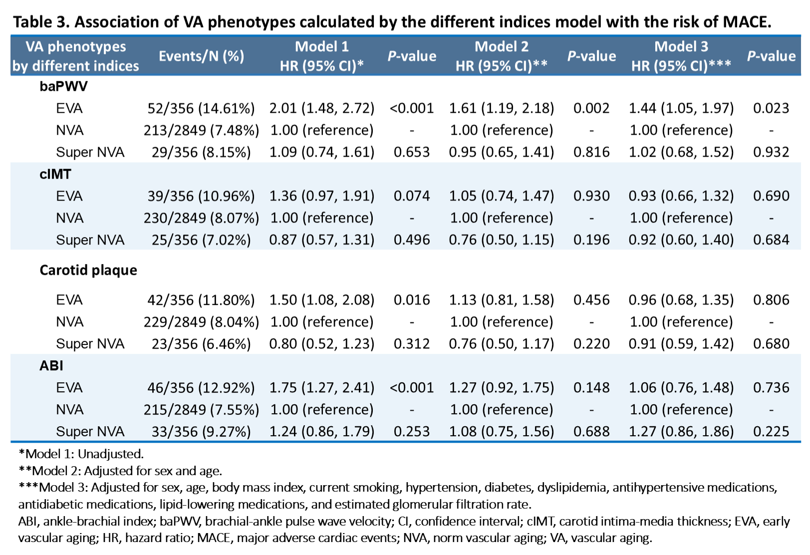
Abstract Body (Do not enter title and authors here): Introduction: Vascular aging (VA) refers to the progressive deterioration of vascular structure and function with age. Vascular age exceeding chronological age indicates higher cardiovascular risk. Indices such as brachial-ankle pulse wave velocity (baPWV), carotid intima-media thickness (cIMT), carotid plaque, and ankle-brachial index (ABI) are often used to assess VA. Research questions: The comparative performance of VA models constructed using these different indicators has not yet been evaluated. This study aimed to compare the cardiovascular risk predictive value of VA models based on baPWV, cIMT, carotid plaque, and ABI. Methods: This study included participants from a Chinese community-based prospective cohort, excluding those with baseline stroke or myocardial infarction. Vascular age was calculated from multivariable regression models incorporating traditional cardiovascular risk factors, medications, and a vascular index (baPWV, cIMT, carotid plaque, or ABI). Residuals between vascular age and chronological age were defined as ΔAge. VA phenotypes were classified as supernormal VA, normal VA (NVA), or early VA (EVA) based on the 10th and 90th percentiles of ΔAge. The endpoint was major adverse cardiovascular events (MACE), including cardiovascular mortality, nonfatal stroke, and nonfatal myocardial infarction. Restricted cubic spline analyses and Cox proportional hazards models were used to examine the association of ΔAge and VA phenotypes with MACE. Results: A total of 3,561 participants (mean chronological age: 58.7±8.5 years) were included. During a mean follow-up of 7.2±1.3 years, 294 (8.26%) participants experienced MACE. The VA model based on baPWV showed the highest explanatory power (R2 = 0.505) (Table 1).ΔAge based on baPWV showed a backwards L-shape association with MACE (Figure 1A). After adjustment, each standard deviation increase in ΔAge based on baPWV was associated with a 14% higher risk of MACE (HR 1.14, 95% CI 1.01–1.27, P=0.027; Table 2). EVA group based on baPWV had a significantly higher risk of MACE compared with NVA group (HR 1.44, 95% CI 1.05–1.97, P=0.023; Table 3). In contrast, VA phenotypes by other indices were not significantly associated with MACE (Table 3). Conclusions: Vascular age based on baPWV demonstrated superior cardiovascular risk prediction versus models using cIMT, plaque, or ABI. baPWV may serve as the preferred method for vascular aging assessment in cardiovascular risk stratification.
Xie, Haotai
(
Peking University First Hospital
, Beijing , China )
Fan, Fangfang
(
Peking University First Hospital
, Beijing , China )
Jia, Jia
(
Peking University First Hospital
, Beijing , China )
Gong, Yanjun
(
Peking University First Hospital
, Beijing , China )
Zhang, Yan
(
Peking University First Hospital
, Beijing , China )
Author Disclosures:
Haotai Xie:DO NOT have relevant financial relationships
| fangfang fan:DO NOT have relevant financial relationships
| JIA JIA:DO NOT have relevant financial relationships
| Yanjun Gong:No Answer
| Yan Zhang:DO NOT have relevant financial relationships
Li Yuxi, Wang Beining, Liu Xu, Zhou Fude, Qiu Jianxing, Lin Zhiyong, Zhou Hui, Ma Dengfeng, Jia Zhi, Xiang Li, Yang Renqiang, Zheng Jingang, Shi Dongmei, Kario Kazuomi, Zhang Yan, Li Jianping, Lu Chengzhi, Fan Fangfang, Liu Zhihao, Liu Shengcong, Yi Tieci, Zhang Long, Weng Haoyu