Empagliflozin Attenuates Endothelial Senescence by Suppressing the Senescence Associated Secretory Phenotype
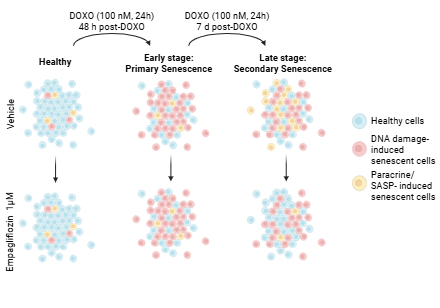
Abstract Body: Background: Endothelial senescence contributes to vascular aging and cardiovascular disease. Senescence is a stress response (e.g., DNA damage, inflammation) in which cells exit the cell cycle and develop a senescence-associated secretory phenotype (SASP). SASP varies with the initiating insult and matures over time: early senescence is dominated by primary, damage-initiated cells with an immature SASP, whereas later, a mature SASP can propagate senescence via paracrine signaling (secondary senescence). SGLT2 inhibitors improve endothelial function, but the molecular mechanisms remain unclear. We hypothesized that empagliflozin (EMPA) mitigates endothelial senescence by regulating SASP. Methods: HUVECs were exposed to doxorubicin (DOXO, 100 nM, 24 h) to induce senescence. To examine the effect of EMPA on early senescence, EMPA (1 µM) was co-incubated with DOXO for 24 h, followed by EMPA alone for an additional 48 h. In late senescence, after DOXO (24 h), cells recovered (4 days), and EMPA was introduced for 72 h. Outcomes included viability and senescence markers, including SA-β-gal, cell-cycle genes (p16, p21, p53), DNA damage (53BP1), telomere dysfunction-induced foci (TIFs), and a SASP profile (Tnfα, IL1α/β, IL6, IL8, Ccl2, Cxcl2, Cxcl10, Serpine1, Mmp3, Gdf15, Tgfb1, Icam, and Vcam). Results: EMPA maintains >80% cell viability across 0.1–100 µM (n=4). DOXO increased SA-β-gal positive cells from ~20% (CTL) to ~80% at both early and late stages, whereas EMPA (1 µM) reduced them to ~60% (n=6; both p<0.05). In early senescence, DOXO increased both p21 and p53 levels, whereas EMPA prevented only the increase in p53 (p=0.005). In late senescence, DOXO increased, and EMPA reduced all three: p16 (~19%; p=0.0008), p21 (~45%; p=0.04), and p53 (~33%; p=0.01). DOXO increased the number of cells with 53BP1 foci (~80% in early, ~60% in late) and TIFs (~40% in early, ~30% in late) (n=3–6), with higher mean foci per cell in early vs late (53BP1: ~5 vs ~3; TIF: ~3 vs ~2). EMPA did not reduce DNA/telomere damage (all p>0.05). DOXO induced SASP at both stages (n=4–6); however, EMPA suppressed SASP predominantly in late senescence (~20–30% reduction across most targets). Notably, Tgfb1 decreased in both early (~22% reduction; p=0.04) and late (~29% reduction; p=0.01) senescence. Conclusions: EMPA attenuates endothelial senescence mainly by suppressing late-stage SASP, with minimal effects on primary DNA/telomere damage, suggesting EMPA limits paracrine/secondary senescence.
Pontes Oliveira De Almeida, Arthur
(
University of Utah
, Salt lake city , Utah , United States )
Virgolino Da Silva Pontes, Larisse
(
University of Utah
, Salt lake city , Utah , United States )
Moreno, Denisse
(
University of Utah
, Salt lake city , Utah , United States )
Housden, Calleen
(
University of Utah
, Salt lake city , Utah , United States )
Lesniewski, Lisa
(
University of Utah and VA Medical center
, Salt lake city , Utah , United States )
Donato, Anthony
(
University of Utah and VA Medical Center
, Salt lake city , Utah , United States )
Author Disclosures:
Arthur Pontes Oliveira de Almeida:DO NOT have relevant financial relationships
| Larisse Virgolino da Silva Pontes:DO NOT have relevant financial relationships
| denisse Moreno:No Answer
| Calleen Housden:No Answer
| Lisa Lesniewski:DO NOT have relevant financial relationships
| Anthony Donato:No Answer