Final ID: P-230
Elevated Blood Pressure Promotes Perivascular Adipocyte Progenitor Proliferation and Alters Their Spatial Distribution
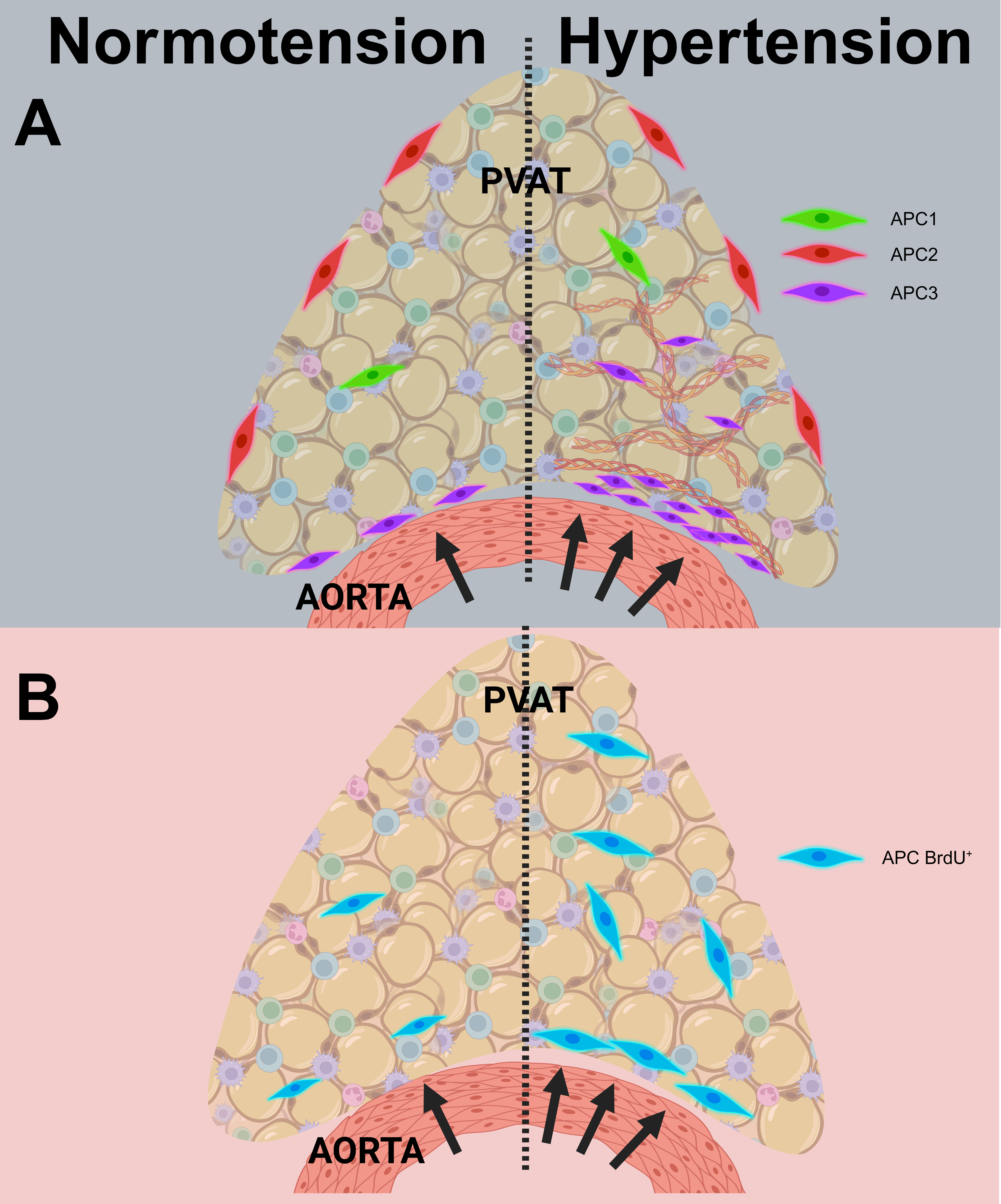
Abstract Body: Perivascular adipose tissue (PVAT), the outermost layer of blood vessels, regulates vascular function. Hypertension (HTN) induces vascular remodeling, a process characterized by fibrosis and cell proliferation. However, this process is poorly understood in PVAT. Adipocytes in PVAT are maintained by continuous adipogenesis of progenitor cells (APC) expressing PDGFRα. Single-cell studies in non-PVAT depots identified 3 APC subtypes that may develop into adipocytes vs fibroblasts and are differentially distributed in AT (ie, parenchyma, fascia). Changes in APC distribution may contribute to fibrosis and consequently vascular remodeling. Currently, the contribution of APC subtypes to HTN-induced PVAT fibrosis is unknown. This study determined HTN effect on APC abundance and phenotype in thoracic PVAT. We hypothesize that elevated blood pressure increases the abundance of profibrotic APC in PVAT. Male mice (PDGFRa-CreERT2-tdTomato n=12) were used to trace APC (PDGFRα+). HTN was induced using 1) aorta coarctation using a ligature (Coarct), creating a hypertensive (Upstream: Up) and hypotensive (Downstream: Down) gradient. Sham animals underwent surgery without ligature. 2) Angiotensin-II (or Saline) infusion. Blood pressure was measured by telemetry under anesthesia or tail cuff. Blood flow velocity was measured with Doppler ultrasound (mm/sec; reported as ratio between Up:Down). APC proliferation was determined by PDGFRα+/BrdU+ colocalization (reported as % of total APC), and APC subtypes’ spatial distribution was evaluated with SABER-FISH. Cxcl14 defined: APC1, Pi16: APC2, and Gdf10: APC3 (reported as % of total nuclei). Coarctation created a ~19mmHg mean arterial pressure gradient across the ligature. Coarct had a faster blood flow ratio (2.05±0.18 vs sham 1.04±0.03). APC3 abundance increased in the parenchyma of coarcted Up (2.7%±1.1) compared to Down (0%); whereas sham animals had no APC3 in the parenchyma of both sites. Similarly, coarct adventitia had more APC3 Up (18.84%± 6.8) than Down (4.5%±2.2). Ang-II increased mean arterial pressure by ~56mmHg compared to saline. APC proliferated in Ang II-treated animals (72%±2.8%) compared to control (45%±8.9%). Our results demonstrate that HTN increased APC abundance, specifically APC3. In conclusion, we identify APC3 as a possible driver of PVAT remodeling during HTN (Figure). Future studies will evaluate how APC subtypes contribute to fibrosis in vitro.
More abstracts on this topic:
Abdominal Aortic Perivascular Adipose Tissue Lipolysis Is Activated In Hypertensive Dahl SS Rats Fed a High-Fat Diet
Chirivi Miguel, Rendon Javier, Lauver Adam, Fink Gregory, Watts Stephanie, Contreras Andres
Chronic Oxidative Stress Induces Hypertension and Abdominal Aortic Aneurysm in Chemogenetic Mice ModelDas Apabrita, Waldeck-weiermair Markus, Yadav Shambhu, Covington Taylor, Spyropoulos Fotios, Pandey Arvind, Michel Thomas

