Large-scale proteomic analysis of valvular calcification: The Atherosclerosis Risk in Communities (ARIC) Study
Abstract Body: Background: Valvular heart disease shares risk factors with atherosclerosis, but its pathophysiology is not fully understood. We aimed to conduct a large-scale proteomic analysis for aortic valve calcification (AVC) and mitral valve calcification (MVC), an early manifestations of valvular heart disease, and compare the results with coronary artery calcification (CAC), a representative marker of atherosclerosis.
Hypothesis: Certain proteins will be uniquely associated with AVC or MVC.
Methods: We first studied 1,704 ARIC participants (mean age 74 [SD 4] years, 61% female, 17% Black), with data on 4,955 plasma proteins (SomaLogic) at Visit 5 (2011-13) and AVC, MVC, and CAC by cardiac CT at Visit 7 (2018-19). We used linear and logistic regression adjusted for traditional risk factors, modeling AVC, MVC, and CAC as continuous (log Agatston score) and as binary (≥75th vs <75th age and sex-specific percentile) outcome variables. Then, we tested whether proteins significant in both models for either AVC or MVC are associated with incident cardiovascular disease (CVD), including coronary heart disease, stroke, and heart failure, using multivariable Cox models. We applied a false discovery rate (FDR) p<0.05 to account for multiple comparisons.
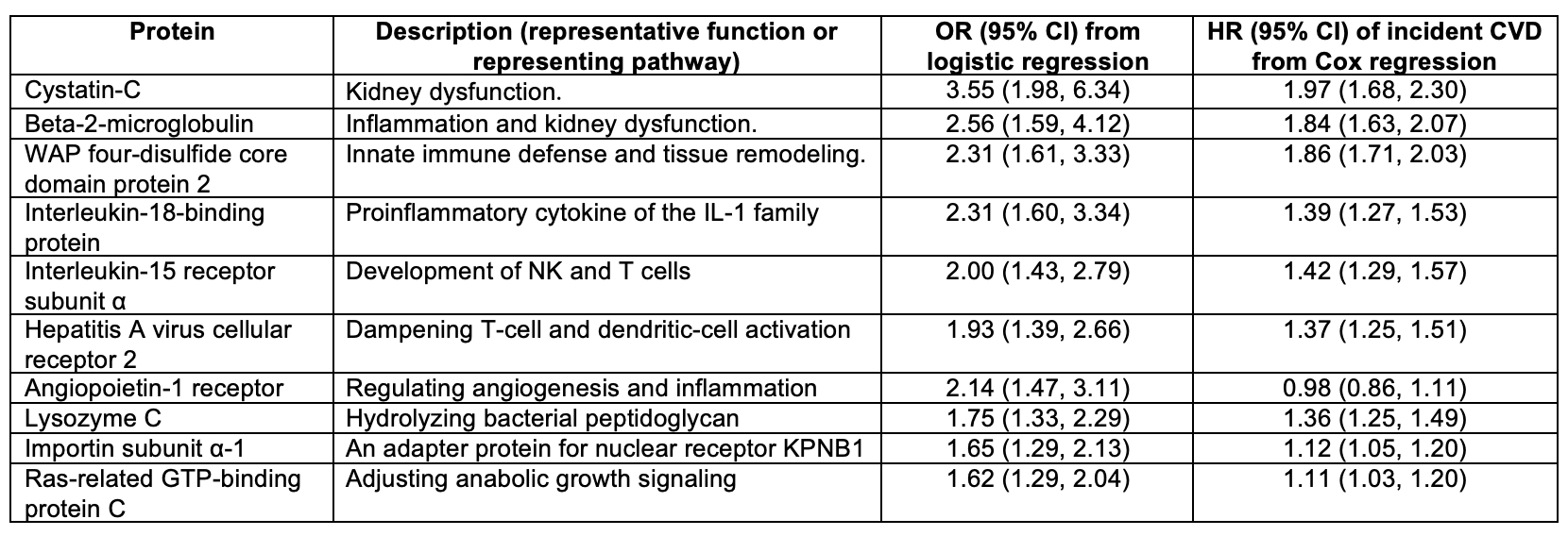
Results: The median (IQR) Agatston score was 0 (0-58) for AVC, 0 (0-115) for MVC, and 244 (38-767) for CAC. For linear regression, 70 proteins were significantly associated with MVC. For AVC, tumor necrosis factor receptor superfamily member 11B (TNFRSF11B) was the only significant protein in linear models, which was one of the 70 proteins associated with MVC. Out of the 70 MVC proteins, 10 were also significant in logistic regression models. WAP four-disulfide core domain protein 2 (WFDC2) and interleukin-18–binding protein (IL-18BP) showed the strongest positive associations in MVC. TNFRSF11B was not significantly associated with AVC in a logistic regression. None of those 10 MVC proteins were associated with CAC. In survival analysis using Visit 2 (1990-92) as baseline, 9 of the 10 MVC proteins were associated with incident CVD. For example, WFDC2 and IL-18BP had hazard ratios of 1.86 (95%CI 1.71-2.03) and 1.39 (1.27-1.53), respectively.
Conclusions: We identified more proteins associated with MVC than AVC, which did not overlap with each other or CAC, suggesting distinct pathophysiology across vascular diseases and atherosclerosis. Notably, most of those MVC proteins were independently associated with future CVD.
Liu, Hairong
(
Johns Hopkins University
, Baltimore , Maryland , United States )
Mok, Yejin
(
Johns Hopkins University
, Baltimore , Maryland , United States )
Yu, Bing
(
UNIV OF TX HEALTH SCI CTR HOUSTON
, Houston , Texas , United States )
Ballantyne, Christie
(
BAYLOR COLLEGE MEDICINE
, Houston , Texas , United States )
Shah, Amil
(
UT Southwestern Medical Center
, Dallas , Texas , United States )
Whelton, Seamus
(
Johns Hopkins School of Medicine
, Baltimore , Maryland , United States )
Blaha, Michael
(
JOHNS HOPKINS HOSPITAL
, Baltimore , Maryland , United States )
Matsushita, Kunihiro
(
JOHNS HOPKINS UNIVERSITY
, Baltimore , Maryland , United States )