
Cardiomyocyte Alignment as a Determinant of Cardiomyocyte Function and Health
Abstract Body: The heart’s ability to efficiently contract relies on the highly ordered alignment of cardiomyocytes across the myocardium. Disruption of this architecture, commonly described as cardiomyocyte disarray (misalignment), is a defining feature of several cardiac diseases and is strongly associated with impaired contractility and disease progression. Despite this clear association between cardiomyocyte disarray and cardiac disease, a fundamental question remains unresolved: does cardiomyocyte misalignment actively contribute to myocardial dysfunction, or does it merely reflect structural remodeling?
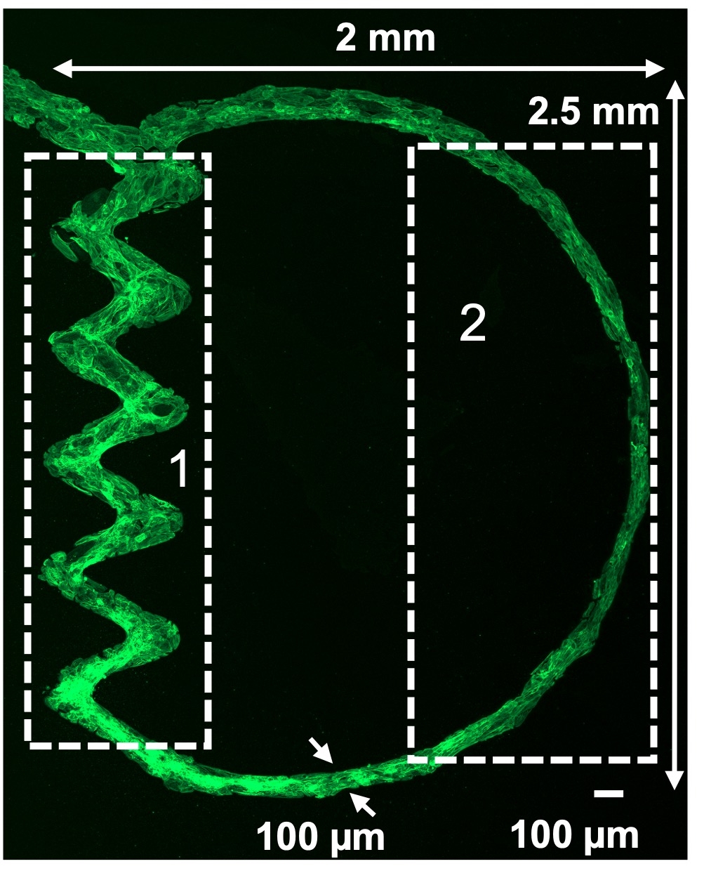
We hypothesize that cardiomyocyte geometric misalignment directly contributes to cardiomyocyte dysfunction, independent of other cellular factors. To test this hypothesis, we developed a controlled experimental system that enables direct comparison of interconnected aligned and misaligned cardiomyocyte regions within the same microenvironment. A 100 µm thin 2D structure resembling the alignment of cardiomyocytes in the mid-myocardium was reproducibly created by seeding hiPSC-derived cardiomyocytes onto predefined Matrigel-based patterns. To mimic a disease condition resembling septal infarction, another structure was created with a misaligned zigzag region representing the infarct zone, connected to a remote region with aligned cardiomyocytes (Fig. 1).
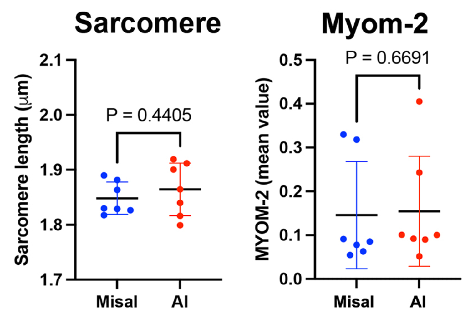
Cardiomyocyte function was evaluated using confocal imaging of calcium handling, mitochondrial function, apoptosis, and autophagy. Cardiomyocytes in the remote region exhibited coherent alignment, whereas cells within the disease-mimicking zigzag region displayed reduced orientation. Importantly, maturation metrics, including sarcomere length and MYOM2 expression, were comparable between aligned and misaligned regions, indicating similar levels of cardiomyocyte maturation and excluding maturation as a contributor to the functional differences observed (Fig. 2).
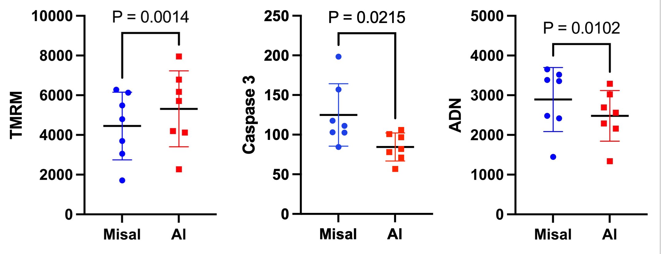
The misaligned region exhibited clear signs of energetic and metabolic stress, including reduced mitochondrial membrane potential, increased autophagic activity, and elevated caspase-3 activity, consistent with greater mitochondrial dysfunction and apoptosis relative to the aligned region (Fig. 3).
Together, these findings suggest that cardiomyocyte misalignment alone can compromise cardiomyocyte health and survival, supporting a potential causal role for architectural disorganization in the pathogenesis of cardiac disease.
Kumar, Alok
(
Massachusetts General Hospital
, Charlestown , Massachusetts , United States )
Mahamdeh, Mohammed
(
Massachusetts General Hospital
, Boston , Massachusetts , United States )
Rock, Christopher Allen
(
Massachusetts General Hospital
, Charlestown , Massachusetts , United States )
Sosnovik, David
(
Massachusetts General Hospital
, Charlestown , Massachusetts , United States )