Second Harmonic Generation Microscopy to Assess Cardiac Fibrosis Changes in a Murine Model of Non-Ischemic Heart Failure
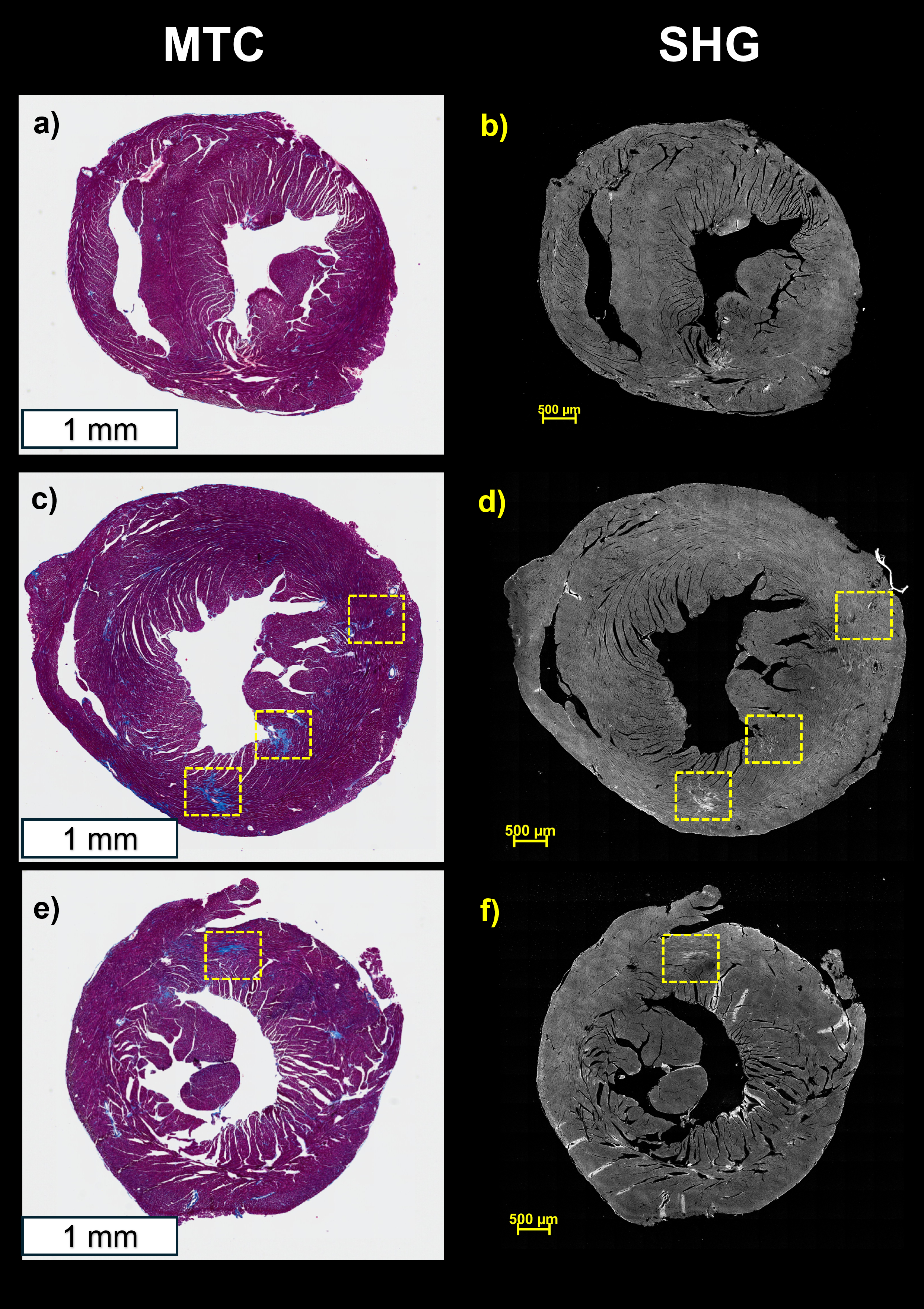
Abstract Body (Do not enter title and authors here): Introduction Heart failure (HF) is characterized by maladaptive changes leading to excessive fibrosis. Delineating the extent of cardiac fibrosis and its specific composition can aid in understanding and development of therapeutics targeting fibrosis. Second harmonic generation (SHG) microscopy, a nonlinear optical technique, enables label-free imaging of collagen remodeling, and provides an opportunity for translation to human applications. In this study, we use whole-slide SHG to assess fibrotic changes in a therapeutic experiment using a non-ischemic mouse model of HF. Hypothesis SHG can characterize fibrotic regions in a non-ischemic mouse model of HF. Methods Non-ischemic HF model was induced in a 3-month-old C57BL6 mouse using drinking water containing 1% NaCl, 0.3 mg/mL L-NAME and continuous infusion of Angiotensin II (0.7 mg/kg/day) using a subcutaneous osmotic pump. In the treatment group, a therapeutic agent was injected (1 mg/kg) subcutaneously twice a week during the HF induction protocol for 5 weeks. Hearts were formalin-fixed, paraffin-embedded, and sectioned (5 μm). Adjacent slides underwent Masson’s Trichrome (MTC) staining. SHG imaging was performed using a femtosecond laser (803 nm, Insight DeepSee, CA) and a Bergamo multiphoton microscope (Thorlabs, NJ) at 25× magnification. An emission filter (400 ± 10 nm) and photomultiplier tube collected backward SHG signals. Whole-slide scans were generated and processed to remove stitching artifacts. Results Both the MTC and the label-free SHG images exhibited an increase in fibrosis in the HF group compared to control, while the fibrosis level was reduced following treatment (Fig. 1). Notably, the SHG images (Fig. 1 b,d,f) did not recapitulate some fibrosis (blue) regions from the brightfield images (Fig. 1 a,c,e), likely due to SHG’s specificity for collagen types I and III, and MTC staining a broader range of collagen types. In all groups, SHG signals were not just localized to the blue regions in the MTC images, but throughout the entire section which could be attributed to SHG originating from myosin in cardiomyocytes. However, fibrosis regions exhibited stronger SHG intensity than myosin, consistent with their higher nonlinear optical signal efficiency. Conclusion SHG microscopy provides high-resolution, label-free detection of changes in cardiac fibrosis, both during progression and its partial reversal with treatment, highlighting its utility for evaluating therapeutic outcomes in HF.
Poon, Wesley
(
Houston Methodist Hospital
, Houston , Texas , United States )
Uzma, Rania
(
Rice University
, Houston , Texas , United States )
Krishnamoorthi, Muthu Kumar
(
Houston Methodist Hospital
, Houston , Texas , United States )
Raghunathan, Raksha
(
Houston Methodist Hospital
, Houston , Texas , United States )
Zhao, Hong
(
Houston Methodist Hospital
, Houston , Texas , United States )
Wong, Stephen
(
Houston Methodist Hospital
, Houston , Texas , United States )
Bhimaraj, Arvind
(
HOUSTON METHODIST HOSPITAL
, Houston , Texas , United States )
Author Disclosures:
Wesley Poon:DO NOT have relevant financial relationships
| Rania Uzma:No Answer
| Muthu Kumar Krishnamoorthi:DO NOT have relevant financial relationships
| Raksha Raghunathan:No Answer
| Hong Zhao:No Answer
| Stephen Wong:DO NOT have relevant financial relationships
| Arvind Bhimaraj:DO have relevant financial relationships
;
Consultant:Abiomed:Active (exists now)
; Advisor:CareDx:Past (completed)
; Research Funding (PI or named investigator):Cardiol Therapeutics:Active (exists now)
; Consultant:Pfizer:Past (completed)
; Speaker:Bridgebio:Past (completed)
; Speaker:Abbott:Active (exists now)