
Ensembled AI Model Can Detect Carotid Artery Intraplaque Hemorrhage from CT Angiograph
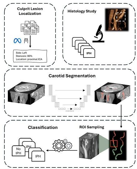
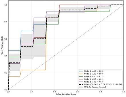
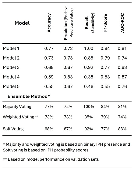
Abstract Body (Do not enter title and authors here): Introduction: Carotid artery intraplaque hemorrhage (IPH) is increasingly recognized as a predictive marker of cerebrovascular events. Despite widespread utilization of contrast-enhanced CT angiography (CTA) for the evaluation of carotid atherosclerosis, diagnosis of IPH on CTA is challenging, limiting risk stratification capabilities. We evaluated convolutional neural networks (CNN) for detection of histology-proven IPH in carotid CTA. Methods: The study included carotid endarterectomy patients from our center who had preoperative CTA and available plaque specimens from the Mayo Clinic Carotid Artery Atherosclerosis Trial (MCAT) Biobank. A validated segmentation model localized common and internal carotid arteries (CCA/ICA). CCA bifurcation served as landmark after a large language model, Llama3.0-70B, verified culprit plaque proximity to the bifurcation from CTA reports. A 3×3×4cm box around ipsilateral CCA bifurcation was sampled from CTA volumes and images were labeled IPH by histology study of carotid specimen (Fig 1). Given limited cases with concurrent imaging and histology, we employed a robust cross-validation strategy: 22 cases (20%) held-out for testing, with remaining data 86 cases used for 5-fold cross-validation (stratified by IPH presence). The five resulting models independently generated IPH probability scores, and ensembled predictions calculated by three methods to enhance generalizability: Majority and weighted voting based on binary IPH class and soft voting based on IPH probability scores. Results Of 418 patients with histological analysis, 130 had CTA studies available. After excluding 22 cases lacking histological consensus, 108 cases were analyzed. In the 22 test cases the mean age was 68.1±8.5 years and 59% were male; In the cross-validation folds age ranged from 67.1±6.9 to 75.3±7.6 years 56-82% were male. Individual models' performance on the test set showed mean ROC-AUC of 0.79 (95% CI: ±0.05) and average precision of 0.85 (±0.05) (Fig 2). The ensemble approach achieved superior performance with soft voting demonstrated 82% ROC-AUC, 77% accuracy, 79% precision, 85% sensitivity, and 82% F1-score for IPH detection, outperforming majority voting and weighted voting (Fig 3). Conclusion: Automated IPH detection in widely available CTA imaging can turn it to a powerful stroke risk assessment tool, supporting personalized prevention strategies and surgical decision-making, ultimately reducing cerebrovascular morbidity and mortality.
Mahmoudi, Elham
(
Mayo Clinic
, Rochester , Minnesota , United States )
Nardi, Valentina
(
Mayo Clinic
, Rochester , Minnesota , United States )
Saba, Luca
(
University of Cagliari
, Cagliari , Italy )
Benson, John
(
Mayo Clinic
, Rochester , Minnesota , United States )
Lerman, Amir
(
Mayo Clinic
, Rochester , Minnesota , United States )
Erickson, Bradley
(
Mayo Clinic
, Rochester , Minnesota , United States )
Author Disclosures:
Elham Mahmoudi:DO NOT have relevant financial relationships
| Valentina Nardi:DO NOT have relevant financial relationships
| Luca Saba:No Answer
| John Benson:DO NOT have relevant financial relationships
| Amir Lerman:DO NOT have relevant financial relationships
| Bradley Erickson:DO NOT have relevant financial relationships