Uncovering Cellular and Molecular Mechanisms in Fontan-Associated Liver Disease
Abstract Body (Do not enter title and authors here): Introduction: Nearly 1 in 10,000 infants are born with a single ventricular heart, and these children must undergo the Fontan procedure to survive. As of 2020, an estimated 50,000 individuals are alive with a Fontan circulation. Although lifesaving, Fontan procedure is associated with several complications, including Fontan-associated liver disease (FALD). While elevated central venous pressure (CVP) appears to promote FALD, the cellular and molecular mechanisms involved in this unique liver disease are not well understood.
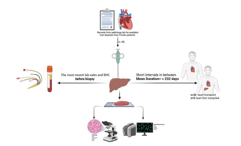
Methods: Liver biopsies from Fontan patients were collected at Washington University in St. Louis before heart transplant. Liver tissue was fixed in formalin and embedded in paraffin for H&E and Masson's trichrome staining. Immunohistochemistry, scRNA-seq, and immunofluorescence (IF) were performed. In our preclinical model, mice underwent pIVCL to mimic hepatic congestion and underwent IF, RNA analysis, and snRNA-seq.
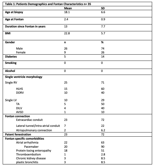
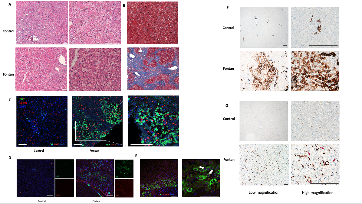
Results: We recruited 35 patients with a mean age of 18.1 years (SD ±6.6) at the time of liver biopsy. The mean age at the time of the Fontan procedure was 2.4 years (SD ±0.9), with the average duration of Fontan circulation being 13 years. Regarding ventricular morphology, 25 patients (71%) had a single right ventricle, and 10 (29%) had a single left ventricle. The extracardiac conduit was the most common Fontan connection in 23 patients (72%). CVP was elevated in the superior vena cava (SVC) and Fontan conduit, with a mean of 18 mmHg (SD ±3.6). Liver function tests showed a median AST of 30.5 U/L (IQR 29.8), median ALT of 26.5 U/L (IQR 26.9), and median total bilirubin of 1.0 mg/dL (IQR 0.9). The mean APRI score was 0.58 (SD ±1.9), and MELD-XI was 10.2 (SD ±2.9). Fontan liver biopsies and liver sections from pIVCL mice showed sinusoidal dilatation, central hepatocyte dropout, and bridging fibrosis. Multiple areas of CK7-positive biliary metaplastic cells, which originated from HNF4α-positive hepatic progenitor cells, were identified. These areas colocalized with areas of CD68-positive aggregated macrophages and activated hepatic stellate cells (αSMA+). Using snRNA-seq in pIVCL and scRNA-seq in Fontan, we identified the cellular landscape in the cardiogenic liver disease.
Conclusions: The interplay between sinusoidal congestion, macrophage activation, and biliary metaplasia in FALD implies a multifactorial pathogenesis that extends beyond passive venous congestion in cardiogenic liver diseases.
Elesawy, Mahmoud
(
WASHINGTON UNIVERSITY IN ST LOUIS
, St Louis , Missouri , United States )
Park, Arick
(
WASHINGTON UNIVERSITY IN ST LOUIS
, St Louis , Missouri , United States )
Fu, Christina
(
WASHINGTON UNIVERSITY IN ST LOUIS
, St Louis , Missouri , United States )
Chan, Mandy
(
WASHINGTON UNIVERSITY IN ST LOUIS
, St Louis , Missouri , United States )
Schilling, Joel
(
WASHINGTON UNIVERSITY IN ST LOUIS
, St Louis , Missouri , United States )
Author Disclosures:
Mahmoud Elesawy:DO NOT have relevant financial relationships
| Arick Park:No Answer
| Christina Fu:DO NOT have relevant financial relationships
| Mandy Chan:DO NOT have relevant financial relationships
| Joel Schilling:DO NOT have relevant financial relationships