Hypoxia Conditioned Extracellular Vesicle Therapy Improves Cardiac Function in a Rat Model of Ischemic Cardiomyopathy
Abstract Body (Do not enter title and authors here): Introduction: Adipose-derived mesenchymal stem cells (AD-MSCs) cultured under hypoxic conditions are known to secrete extracellular vesicles (EVs) with protective effects against ischemia. In this study, we aimed to evaluate the enhanced therapeutic potential of these EVs—emerging as a promising cell-free strategy for cardiac regeneration—when derived from hypoxia-conditioned AD-MSCs.
Materials and Methods: MSCs were isolated from the adipose tissue of Lewis rats and cultured under normoxic (20% O2) or hypoxic (5% O2) conditions. EVs were collected from conditioned media via ultracentrifugation. EV marker protein expression assessed the quantity of EVs, and comprehensive miRNA profiling was performed. To evaluate therapeutic efficacy, myocardial infarction (MI) was induced in Lewis rats via ligation of the left anterior descending artery. Two weeks after MI, rats were divided into three groups: normoxic EVs (N-EV), hypoxic EVs (H-EV), and PBS control. Each treatment was locally injected around the infarct border zone. Four weeks later, cardiac function was assessed by echocardiography and histological analyses were conducted.
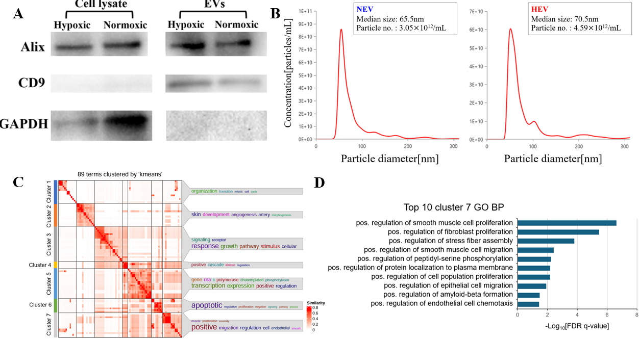
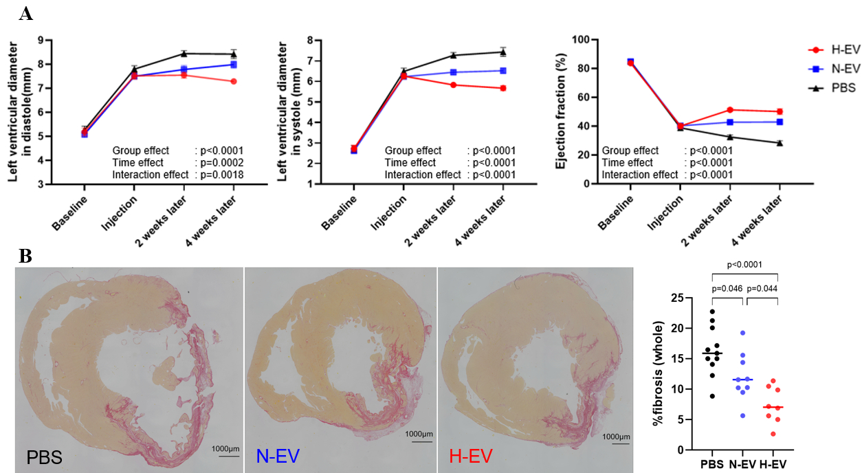
Results: There was no significant difference in the amount of EVs, as assessed by Alix quantification, between the hypoxic-and normoxic groups (p = 0.25)(Figure 1A). There were no significant differences in the median particle size (70.5nm vs 65.5nm, p = 0.51) or concentration (4.59×10¹²/mL vs 3.05×10¹²/mL, p = 0.70) of EVs between the hypoxic and normoxic groups. (Figure 1B). miRNA sequencing revealed that hypoxia-derived EVs contained more miRNAs associated with anti-fibrotic gene regulation (Figure 1C and 1D). In the rat ICM (Ischemic Cardiomyopathy) model, echocardiographic assessment at 4 weeks post-administration revealed that the H-EV group exhibited significantly greater prevention of left ventricular diastolic diameter enlargement (7.3mm vs 8.0mm, p = 0.004) and systolic diameter enlargement (5.7mm vs 6.5mm, p = 0.0004), as well as improved ejection fraction (50% vs 43%, p = 0.0069), compared to the N-EV group (Figure 2A). Histologically, Picro-Sirius Red staining demonstrated a greater reduction in myocardial fibrosis in the H-EV group (7.3% vs 12.0%, p=0.044) (Figure 2B).
Conclusions: In a rat model of ICM, EVs derived from hypoxic MSC cultures were enriched in anti-fibrotic miRNAs and exhibited superior therapeutic effects in terms of remodeling suppression and functional recovery compared to normoxic EVs.
Kumagai, Kunitaka
(
Tottori university Hospital
, Yonago city , Japan )
Kawamura, Takuji
(
Osaka University
, Osaka , Japan )
Torigata, Kosuke
(
Osaka University
, Toyonaka Osaka , Japan )
Harada, Akima
(
Osaka University
, Osaka , Japan )
Miyagawa, Shigeru
(
Osaka University
, Suita , Japan )
Yoshikawa, Yasushi
(
TOTTORI UNIVERSITY
, Yonago,Tottori , Japan )
Author Disclosures:
Kunitaka Kumagai:DO NOT have relevant financial relationships
| Takuji Kawamura:DO NOT have relevant financial relationships
| Kosuke Torigata:No Answer
| Akima Harada:No Answer
| Shigeru Miyagawa:No Answer
| Yasushi Yoshikawa:DO NOT have relevant financial relationships