Left ventricular 3D-sphericity is associated with 92 genetic loci
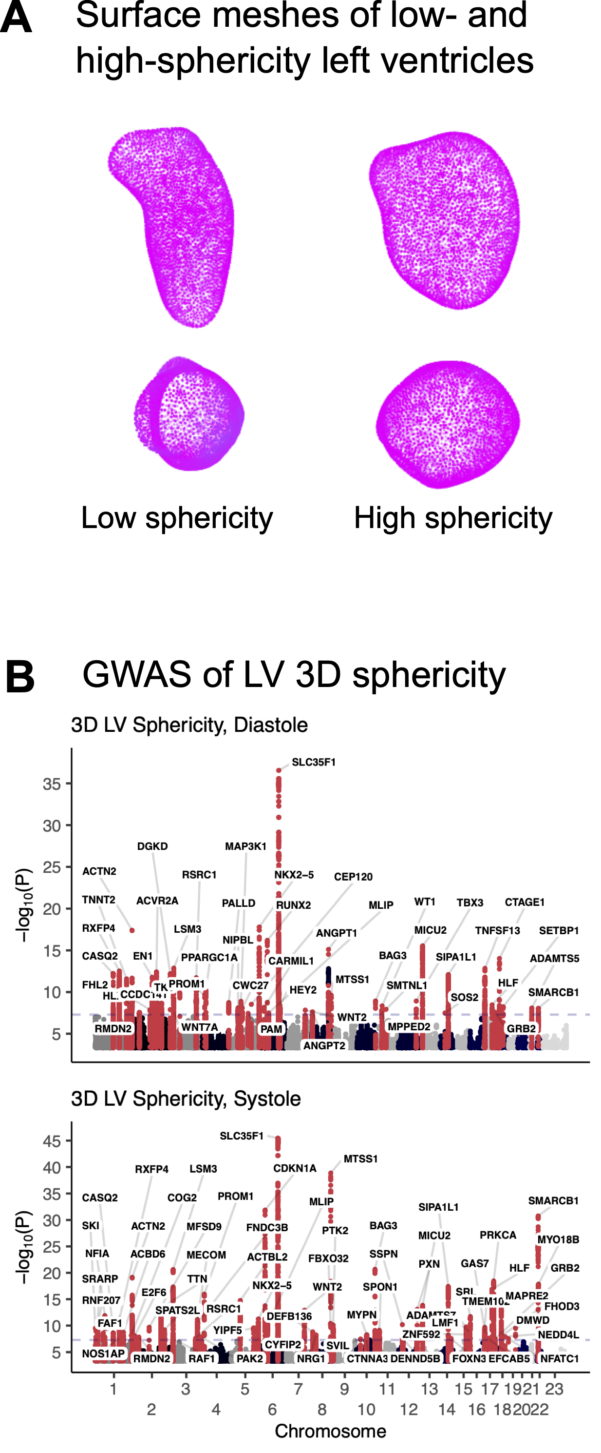
Abstract Body (Do not enter title and authors here): Introduction Left ventricular (LV) sphericity is a measure of the degree to which the LV blood pool resembles a sphere. Analyses of cardiovascular magnetic resonance imaging (MRI) in the MESA cohort have shown that LV sphericity is an important mode of variation in cardiac structure that is associated with ischemia, heart failure, and atrial fibrillation. A recent genome-wide association study (GWAS) of a two-dimensional estimate of sphericity in 38,000 UK Biobank participants identified 4 genetic loci (near PDZRN3, HLA, PLN, and ANGPT1). Genetic contributions to three-dimensional (3D) sphericity remain unknown.
Aims To measure 3D sphericity from cardiovascular MRI at scale and characterize the genetic contributions to 3D sphericity.
Methods Deep learning models were constructed to perform semantic segmentation in the short- and long-axis view from cardiovascular MRI in UK Biobank. These models were applied to all participants with MRI. Poisson surface reconstruction was used to build 3D meshes of the left ventricle. Sphericity was estimated at end-systole and end-diastole as the area of a perfect sphere with the same volume as the left ventricle, divided by the estimated surface area from the mesh. Heritability was estimated with ldsc; GWAS was conducted with REGENIE.
Results Sphericity was estimated in 68,978 individuals, of whom 3,096 were excluded due to atrial fibrillation or atrial dysfunction, and 2,686 were excluded during genetic quality control; 63,196 participants were included in the GWAS. Ldsc heritability was 29.7% for sphericity in diastole and 26.0% for sphericity in systole. 49 genetic loci were associated with 3D sphericity in diastole as were 64 in systole (92 distinct loci). 46 of these loci were unique to sphericity and not found for other LV traits, including loci near the genes encoding calsequestrin 2, troponin T2, palladin, supervillin, and angiopoietins 1 and 2. Shared loci included those near RXFP4, encoding a relaxin-family peptide receptor, and MICU2 (previously implicated in cardiac relaxation).
Conclusion Left ventricular sphericity is heritable, with genetic underpinnings distinct from standard LV measurements.
Pirruccello, James
(
UCSF Division of Cardiology
, San Francisco , California , United States )
Author Disclosures:
James Pirruccello:DO have relevant financial relationships
;
Other (please indicate in the box next to the company name):JACC (Associate Editor):Active (exists now)